


3D Bioprinting Pushes Bone Scaffold Engineering Forward
Bone tissue engineering (BTE) has emerged as a critical solution for repairing large bone defects caused by trauma, infection, or tumors. Traditional grafting techniques—autologous or allogeneic—often face donor shortages, immune rejection, and the need for secondary surgeries. BTE circumvents these issues by seeding osteoblasts or stem cells onto biocompatible, biodegradable scaffolds, which act as a temporary habitat, supporting nutrient exchange and waste removal until new bone forms.

The choice of scaffold material is central to success. Inorganic bioceramics such as hydroxyapatite (HA), tricalcium phosphate (TCP), biphasic calcium phosphate (BCP), and silicate ceramics offer excellent biocompatibility and osteoconductivity. HA’s chemical similarity to natural bone promotes strong bonding, while ?-TCP degrades rapidly, releasing calcium and phosphorus to stimulate regeneration. BCP balances HA’s stability with TCP’s degradability, and silicate ceramics contribute silicon ions that enhance early calcification. Yet, brittleness and slow degradation remain challenges.
Metallic materials, notably magnesium and titanium, provide high mechanical strength. Magnesium’s density and elastic modulus closely match bone, and it degrades into ions safely metabolized by the body. Titanium excels in corrosion resistance and biocompatibility, bonding tightly with host bone. However, metals risk stress shielding and chronic inflammation over long-term implantation.
Organic polymers, both natural and synthetic, bring tunable properties. Natural options like collagen and chitosan support cell adhesion but suffer from poor mechanical strength and variable degradation. Synthetic biodegradable polyesters such as PLA, PGA, PLGA, and PCL allow control over mechanical performance and degradation rates, and have FDA approval for biomedical use. Their acidic degradation byproducts can hinder healing, prompting composite designs that blend polymers with bioceramics to buffer pH and improve osteoinductivity.
Composite scaffolds combine strengths: inorganic components add bioactivity and mechanical stability, while polymers improve toughness and tailor degradation. Ratios can be adjusted to match patient-specific regeneration rates—fast-degrading polymers for rapid healing, slower ceramics for extended support. In high-load regions like the spine, metal-ceramic blends maintain structural integrity; in low-load facial bones, lightweight bioactive composites accelerate integration.
Advanced composites mimic the extracellular matrix (ECM), using porous architectures to promote vascularization and cell migration. Surface functionalization and embedded growth factors further stimulate osteogenesis. Carbon nanotubes (CNTs) have recently been incorporated, enhancing mechanical strength, surface roughness for cell adhesion, and even electrical conductivity for nerve or muscle regeneration.
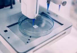
3D bioprinting has revolutionized scaffold fabrication. Using CT and MRI imaging, patient-specific defect geometries are captured and converted via CAD into precise print instructions. Bio-inks—mixtures of cells and biomaterials—are deposited layer-by-layer, enabling complex architectures with controlled porosity and tailored mechanical properties. Low-temperature printing has produced hierarchically porous scaffolds with superior biomineralization.
Printing methods include inkjet, extrusion, and laser-assisted bioprinting, each suited to different material viscosities and resolutions. Scaling production remains a hurdle: maintaining precision, bio-ink consistency, and cell viability across batches is difficult. Multi-nozzle systems and automated material handling are under development to address these limitations.
Applications span critical-sized defect repair, fracture stabilization, craniofacial reconstruction, and even post-tumor resection. For example, magnesium oxide/PLGA scaffolds printed at low temperatures can release magnesium ions and reactive oxygen species to inhibit tumor recurrence while promoting regeneration. Multifunctional designs integrate antimicrobial agents, osteogenic factors, and angiogenic components into a single scaffold.
Design parameters such as porosity (ideally 60–80%), pore size distribution, and gradient degradation profiles are tuned to balance mechanical support with biological integration. Outer layers may degrade quickly to encourage vascularization, while inner layers persist longer for load-bearing support. Immune compatibility is addressed through low-immunogenic materials, bioactive coatings, and pore architectures that facilitate tissue infiltration.
Future directions emphasize developing novel biodegradable composites, integrating imaging for real-time monitoring of scaffold degradation and tissue growth, and refining printing methods for diverse implant environments. As 3D bioprinting technology matures, it promises to deliver patient-specific, multifunctional scaffolds that synchronize mechanical performance with biological healing, marking a significant advance in regenerative medicine.